
Case Presentation
A 26-year-old female, normally healthy and on no medications except oral contraceptive pills, presented to the Emergency Department with:
- 1-day history of left-sided chest pain
- Pain was dull, worse with inspiration
- No clear relation to movement or position
- No associated symptoms
- Vital signs were completely normal
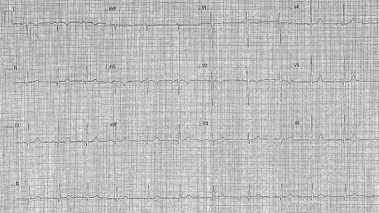
Here is her ECG,
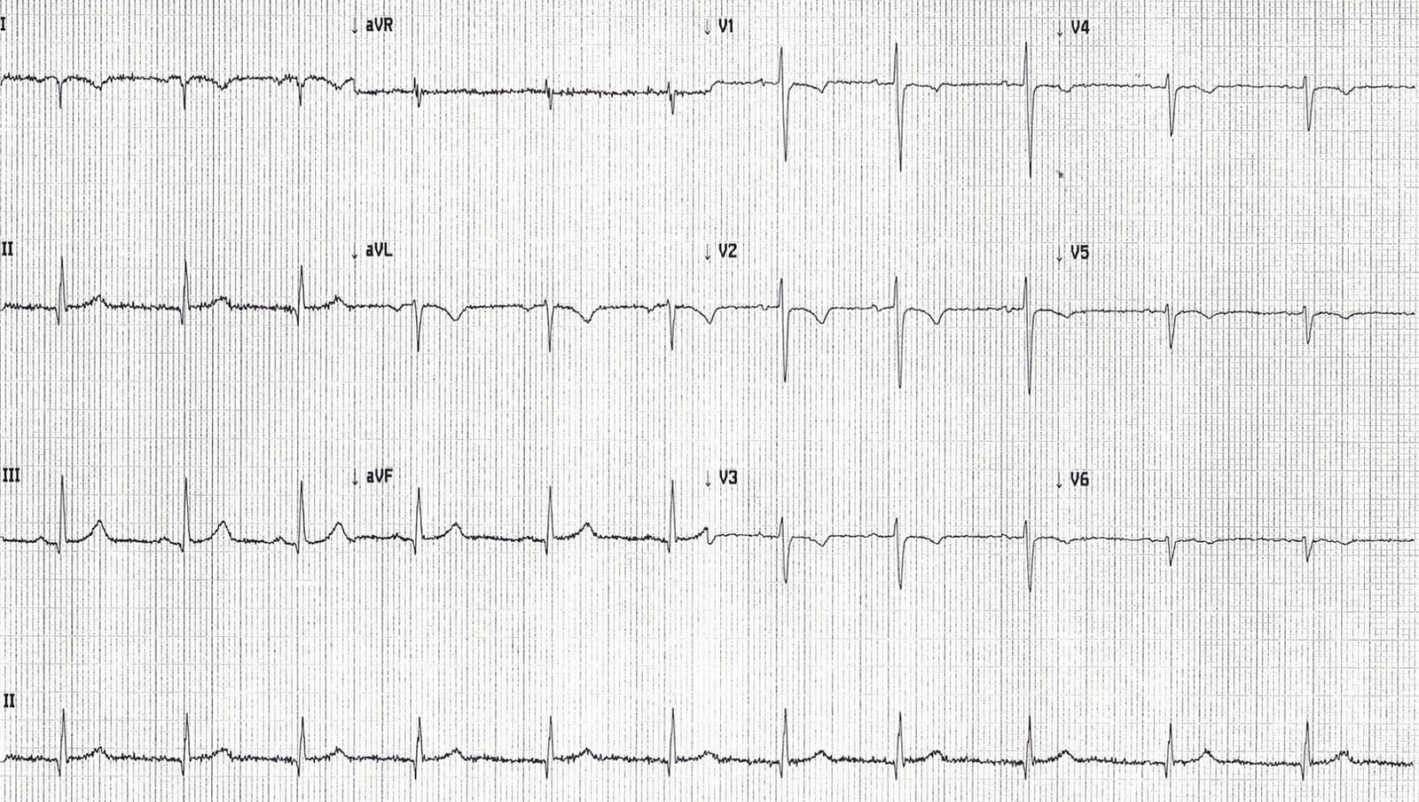
ECG Key Findings:
P waves, QRS complexes, and T waves were negative in lead I and aVL
P wave was positive in aVR
These features raise two important differentials:
1. Limb Lead Malposition
2. Dextrocardia
The ECG shown demonstrated a pattern consistent with limb lead reversal, but is that the full story?
ECG Interpretation Clues
In limb lead malposition, chest leads remain normal because the issue is isolated to limb lead misplacement.
Like in this example:
Additional Abnormalities
- Reversed R wave progression in chest leads (R wave starts tall in V1 and gets smaller toward V6)
- T wave inversion across the chest leads
- Progressively smaller QRS complexes from V1 to V6, suggesting that the electrodes are moving away from the cardiac mass
When you combine:
- Limb lead reversal pattern
- Abnormal chest lead progression (reversed R wave progression).
- Inverted T waves in precordial leads
- Decreasing QRS amplitude across V1–V6
✅ The most likely diagnosis becomes Dextrocardia
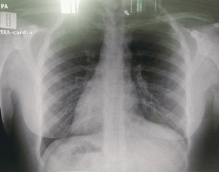
This patient had Dextrocardia with situs inversus totalis,
confirmed by Chest X-ray showing:
- Gastric gas bubble on the right
- Liver shadow on the left
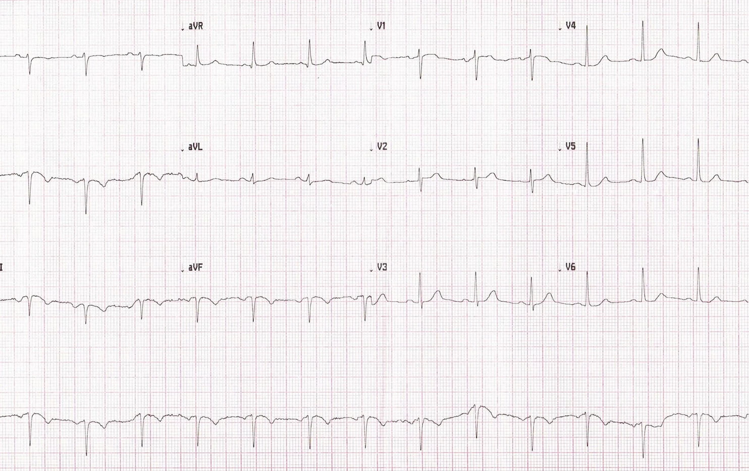
Her ECG below is with the leads placed on the right side of the precodrium
(all abnormalities disapeared)
Final Diagnosis and Disposition
- Patient had low pre-test probability for Pulmonary Embolism (PE)
- D-dimer was negative
- She was discharged with the diagnosis of non-specific chest pain, likely costochondritis
Key Takeaways
- Dextrocardia should be suspected when limb leads show reversed polarity and chest leads have reversed R wave progression
- Limb lead malposition affects only limb leads; chest leads stay normal
- A right-sided ECG is key to confirming true dextrocardia
- Always consider anatomical variations in young patients with chest pain and unusual ECGs
✅ FAQ - Lead Malposition vs Dextrocardia
Q1: How can you differentiate limb lead reversal from dextrocardia on ECG?
A1: Limb lead reversal affects limb leads only, while dextrocardia also causes reversed R wave progression and abnormal chest lead patterns.
Q2: What are signs of dextrocardia on ECG?
A2: Negative P/QRS/T waves in lead I and aVL, reversed R wave progression from V1–V6, and progressively smaller QRS complexes in precordial leads.
Q3: How is dextrocardia confirmed clinically?
A3: With a chest X-ray showing situs inversus and/or a right-sided ECG that normalizes the waveforms.